

What is Retinal Imaging?
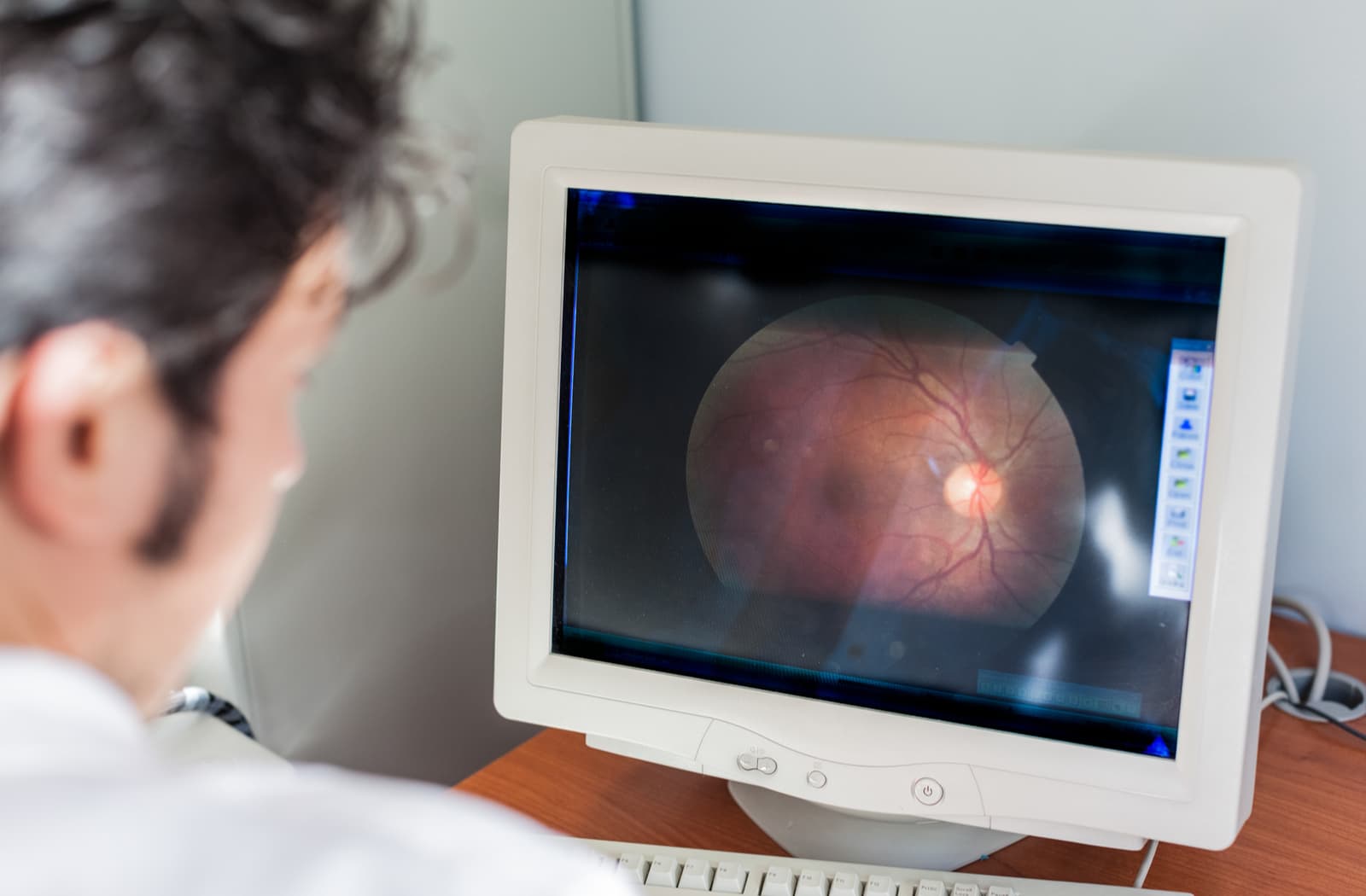
Retinal imaging is a non-invasive diagnostic tool used to take high definition images of the back of your eye. The technology used includes specialized cameras and scanners to magnify your retina, optic nerve, and blood vessels inside the eye.
Your eye’s internal workings are responsible for your vision, so it’s essential to take care of them and properly monitor your health to prevent disease. Typically, the earlier signs of eye disease are caught, the better chance you have of protecting your eyesight.
Different Types of Retinal Imaging
Optical Coherence Tomography (OCT)
Optical coherence tomography is an imaging technology that takes high-resolution images of your retina. This imaging technique takes cross-sectional photos, allowing your optometrist to examine the layers within the retina.
OCT uses rays of light to measure the retinal thickness and is considered the standard of care for assessing and treating most retinal conditions. Because OCT uses beams of light, no radiation or x-rays are used, making it a generally comfortable and painless test.
Optomap Ultra-Widefield Imaging
Optomap ultra-widefield imaging is a diagnostic tool that uses low power lasers to create wide-angle images of your eye. The technology scans your retina to capture up to a 200-degree view of the back of your eye.
Compared to other imaging technologies, which typically only capture 45 degrees, Optomap’s wide angles give your optometrist a great advantage to detect and diagnose early signs of disease. You also don’t need to have your pupils dilated to get such a wide-angle shot of your retina so you can return to normal daily activities following retinal imaging.
Fundus Photography

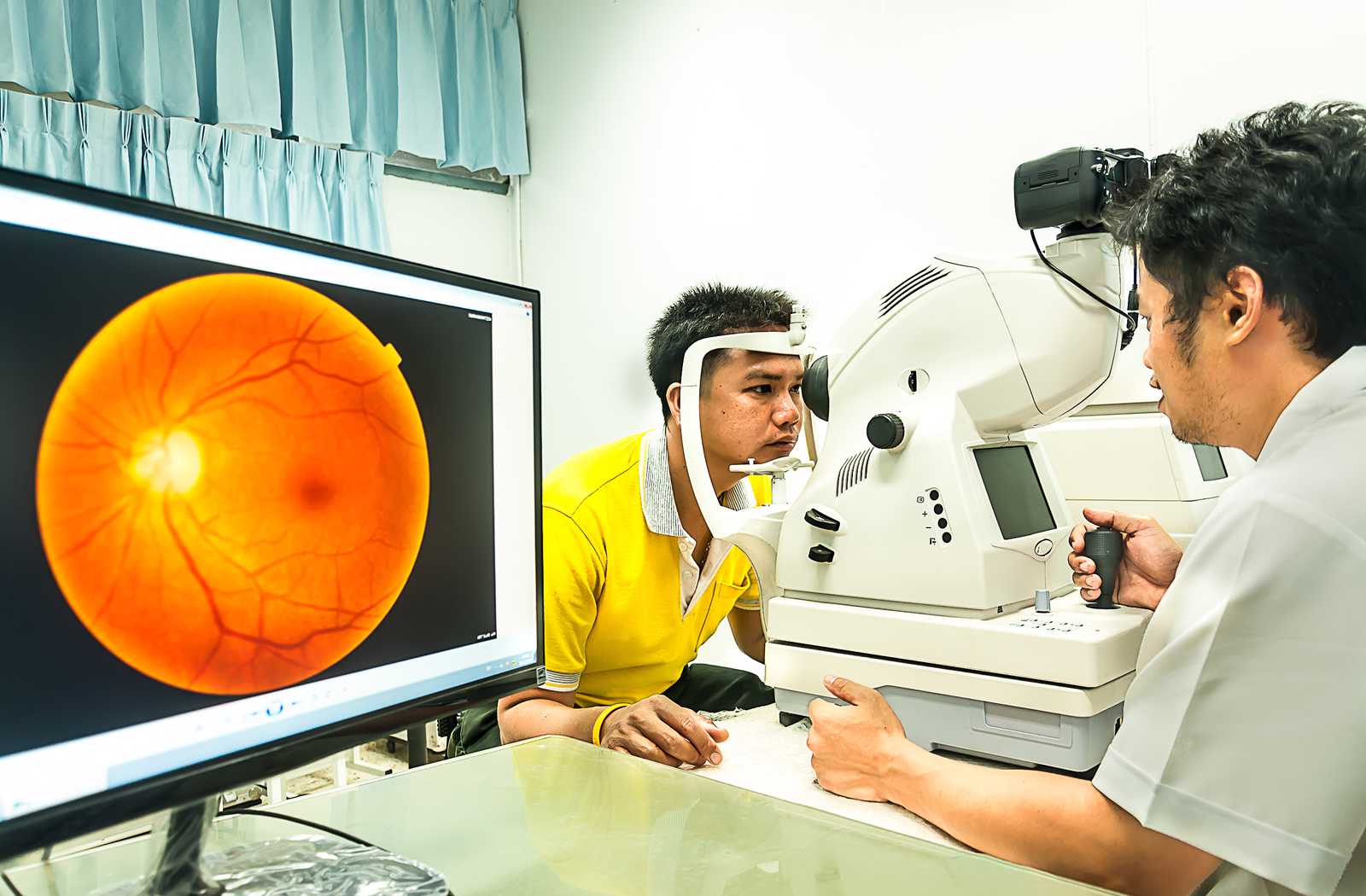
Fundus photography takes high-resolution images of the fundus or the back of the eye. A fundus camera is a specialized, low-power microscope with an attached camera. The procedure typically only takes a minute or two and is generally said to be painless.
The images produced are typically in full colour, which allows your optometrist to quickly identify blood buildup anywhere between the retina and optic nerve. It can also show drusen, scar tissue, and any other abnormalities that indicate early signs of disease.
What Can Retinal Imaging Help Diagnose?
Retinal imaging is done to help detect and diagnose eye problems and monitor their progression or treatment efficacy. At Okotoks Eyecare, retinal imaging is incorporated into a comprehensive eye exam to ensure any early signs of eye disease are caught before they have a chance to worsen.
Early detection of eye disease maximizes your chances of protecting your vision and ocular health.
Glaucoma
Glaucoma is a progressive condition that is often referred to as the “silent thief of sight” because it usually develops without symptoms. As one of the leading causes of blindness in Canada, glaucoma can quickly lead to irreversible vision loss without prompt intervention and treatment.
One of the most common glaucoma causes is elevated intraocular pressure, which often occurs due to a buildup of fluid in the eye. The pressure exerts stress on the optic nerve and damages it, which causes vision loss.
Using retinal imaging technology to view the optic nerve in tandem with visual field testing can detect early signs of glaucoma. Although optic nerve damage cannot be reversed, proper monitoring combined with functional testing and early intervention is imperative in managing glaucoma and protecting your eyesight.
Diabetic Eye Disease
Diabetes is a chronic disease that affects how your body produces and uses insulin, which leads to increased glucose levels in your bloodstream or high blood sugar. Diabetes can also cause several complications that can lead to vision-threatening eye diseases.
Diabetic retinopathy is one of the most common diabetes-related eye diseases. It occurs when the tiny blood vessels in the eye swell and rupture, leaking blood and other fluid into the eye. In advanced stages, the decreased circulation of blood vessels deprives the retina of oxygen, leading to parts of the retina dying off.
If left untreated, diabetic retinopathy can lead to permanent vision loss and even blindness. Retinal imaging is useful in detecting and diagnosing early signs of diabetic eye disease.
Optical coherence tomography and fundus photography help your optometrist get a closer look at the back of your eye. These imaging tests help determine if your blood vessels are normal and if the fluid has begun to leak into your retinal tissue.
Age-Related Macular Degeneration
Age-related macular degeneration (AMD) is a condition that occurs when the centre of the retina begins to deteriorate, causing central vision to become obstructed or blurred. Typically, AMD happens due to the ageing process and is the leading cause of blindness in adults over 55 in North America.
There are 2 main types of AMD, dry and wet. Dry AMD is the more common type, causing a gradual degeneration of the central retinal tissues. Wet AMD happens much more rapidly due to a sudden leakage from weak blood vessels under the macula.
Both optical coherence tomography and fundus photography can be used to pinpoint early signs of age-related macular degeneration. The images produced from both methods can show fluid deposits in the retina, a buildup of drusen, an early sign of dry AMD, scar tissue, and other abnormalities.
Early detection of AMD can help protect your eye health and maximize your chances of saving your vision.
Hypertensive Retinopathy
Hypertensive retinopathy happens as a result of high blood pressure or hypertension. When your blood pressure is too high, the eye’s blood vessels’ walls can thicken and cause the vessels to narrow. Blood vessels that become too narrow restrict blood from reaching the retina, causing the retina to swell. The damage sustained to the blood vessels can limit the retina’s function and pressure the optic nerve, which is responsible for clear vision.
Prolonged high blood pressure is the leading cause of hypertensive retinopathy. The force of your blood pumping from your heart to your arteries is too high, leading to problems over time.
Retinal imaging is imperative in diagnosing hypertensive retinopathy before it damages your vision. The high-resolution images allow your optometrist to evaluate the blood vessels in your eyes, retina, and optic nerve for any abnormalities that indicate early signs of disease.
Your friendly optometrists at Okotoks Eyecare are dedicated to protecting and preserving your eye health and vision. Visit us today for a comprehensive eye exam and find out what you can do to prolong good eyesight.

